

.png)
.png)

Objective: The aim of this study was to detect the relationship between the development of Schmorl’s nodes (SNs) and bone mineral density (BMD) in young patients.
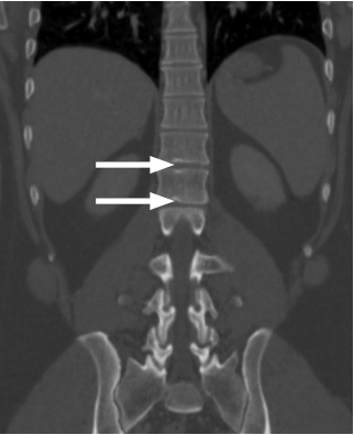
Methods: Computerized tomography (CT) images of the thoracolumbar vertebral column were retrospectively examined by two experienced radiologists for SNs. The diagnostic criterion for SN was defined as a node size larger than one-third but not more than two-thirds of the relevant vertebral endplate. Considering the eligibility criteria, a total of 74 individuals (60 males and 14 females; mean age: 24.3 years; age range: 18-40 years) with SN at the thoracolumbar vertebrae were included in the patient group, and a total of 38 age- and gender-matched individuals (30 males and 8 females; mean age: 25 years) with no evidence of SN were included in the control group. All these individuals were younger than 40 years. In the patient group, SNs were assessed in terms of the distribution of the thoracolumbar vertebrae, the location of the upper and lower endplates, and the total number of lesions. In all individuals included in the study, BMD was measured from the axial CT sections by quantitative CT and then compared between the two groups.
Results: The distribution of age and gender was comparable between the two groups (p=0.438). A total of 208 SNs were identified in the patient group. Of these, 92 (44%) were located at the thoracic vertebrae and 116 (56%) at the lumbar vertebrae. The mean BMD was 131.6 g/cm3 in the patient group and 140.7 g/cm3 in the control group (p=0.03). There was no significant relationship between the total number of SNs per patient and the mean BMD (p=0.156).
Conclusion: Evidence from this study revealed that low BMD may be a predisposing factor for the development of SNs in patients younger than 40 years.
Level of Evidence: Level III, Diagnostic Study
Cite this article as: Güngör Ö, Gezer NS, Özdamarlar U, Balcı A. The effect of bone mineral density on development of Schmorl’s nodes in young patients. Acta Orthop Traumatol Turc 2020; 54(3): 287-92. DOI: 10.5152/j.aott.2020.03.577.