

.png)
.png)

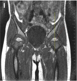
Objective: The aim of this study was to measure the cartilaginous coverage of the acetabulum using magnetic resonance imaging (MRI) and to analyze its effect on the timing and necessity of secondary operations in residual acetabular dysplasia (RAD).
Methods: The MRI results of 33 children (30 girls and 3 boys) aged between 5 and 9 years who were operated on unilaterally via a posteromedial limited approach were compared with the radiographical findings of acetabular dysplasia at follow-up. The acetabular index (AI) and the center-edge (CE) angles were measured. MRI was used to measure the osseous acetabular index (OAI), cartilage acetabular index (CAI), and cartilaginous center-edge angles (CCE). The Children's Hospital's Oakland Hip Evaluation Score (CHOHES) was used for the assessment of clinical and functional results. The Severin scoring system was used to evaluate the radiographic results. The Mann-Whitney U test and Spearman correlation tests were used for statistical analysis.
Results: In all, 30 (90.9%) girls and 3 (9.1%) boys with an average age of 7.4 years (range: 5e9 years) and a mean follow-up period of 6.1 years (range: 4e8 years) were included. While there was a significant difference between non-dislocated hips and operated hips in 3 measurements (AI, Wiberg CE, and Ogata CE) using X-rays (p < 0.05), no significant difference was found in the MRI measurements (OAI, CAI, and CCE) (p > 0.05). The CAI values were lower than the AI measured on X-ray (p ¼ 0.035). The mean CCE was higher than the mean CE (p ¼ 0.022). The mean CHOHES score was 83.1 (range: 52e100) and the score of 62% patients was above 90. There was no significant difference in terms of CHOHES score according to age at the time of operation (p ¼ 0.43). Three (9.1%) patients were Severin class I, 8 (24.3%) patients were class II, 12 (36.3%) patients were class III and 10 (30.3%) patients were class IV. There was no correlation between preoperative hip dislocation and Severin score (p ¼ 0.056). No significant difference was found between the ambulatory and non-ambulatory groups in terms of Severin classification (p ¼ 0.063).
Conclusion: Cartilaginous acetabulum should be taken into account in RAD measurements. MRI may be a more appropriate option for the evaluation of acetabular cartilaginous coverage in the evaluation of RAD and the decision to perform surgery, though X-rays are currently the most used method. The results revealed no effect on functional or radiological scores as a result of being of walking age.
Level of study: Level III, Diagnostic Study.